Cells, Free Full-Text
Por um escritor misterioso
Descrição
High-resolution 3D images of organelles are of paramount importance in cellular biology. Although light microscopy and transmission electron microscopy (TEM) have provided the standard for imaging cellular structures, they cannot provide 3D images. However, recent technological advances such as serial block-face scanning electron microscopy (SBF-SEM) and focused ion beam scanning electron microscopy (FIB-SEM) provide the tools to create 3D images for the ultrastructural analysis of organelles. Here, we describe a standardized protocol using the visualization software, Amira, to quantify organelle morphologies in 3D, thereby providing accurate and reproducible measurements of these cellular substructures. We demonstrate applications of SBF-SEM and Amira to quantify mitochondria and endoplasmic reticulum (ER) structures.

Five-Year Outcomes for Refractory B-Cell Lymphomas with CAR T-Cell Therapy

Streptomyces cell-free systems for natural product discovery and engineering - ScienceDirect

Cells, Free Full-Text
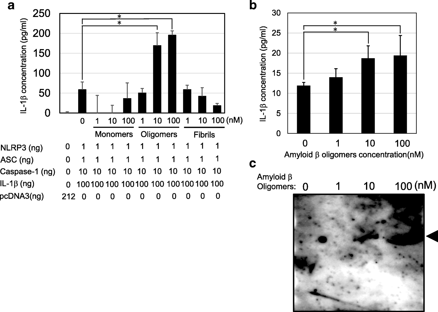
Amyloid β directly interacts with NLRP3 to initiate inflammasome activation: identification of an intrinsic NLRP3 ligand in a cell-free system, Inflammation and Regeneration

Circulating Tumor Cells, Disease Progression, and Survival in Metastatic Breast Cancer
Full-spectrum cell-free RAN for 6G systems: s
Cells, Free Full-Text
Cells, Free Full-Text
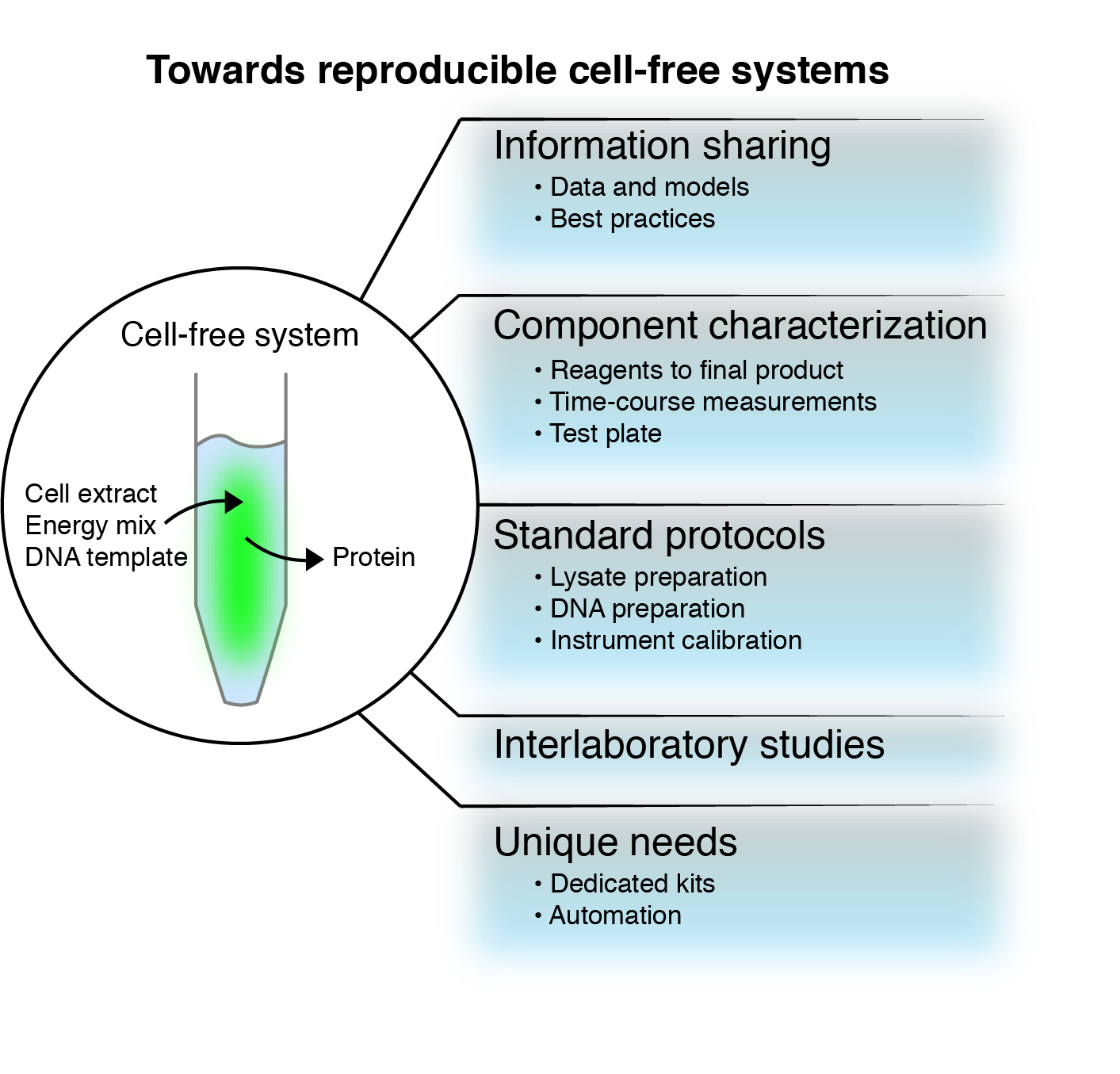
Towards reproducible cell-free systems
de
por adulto (o preço varia de acordo com o tamanho do grupo)