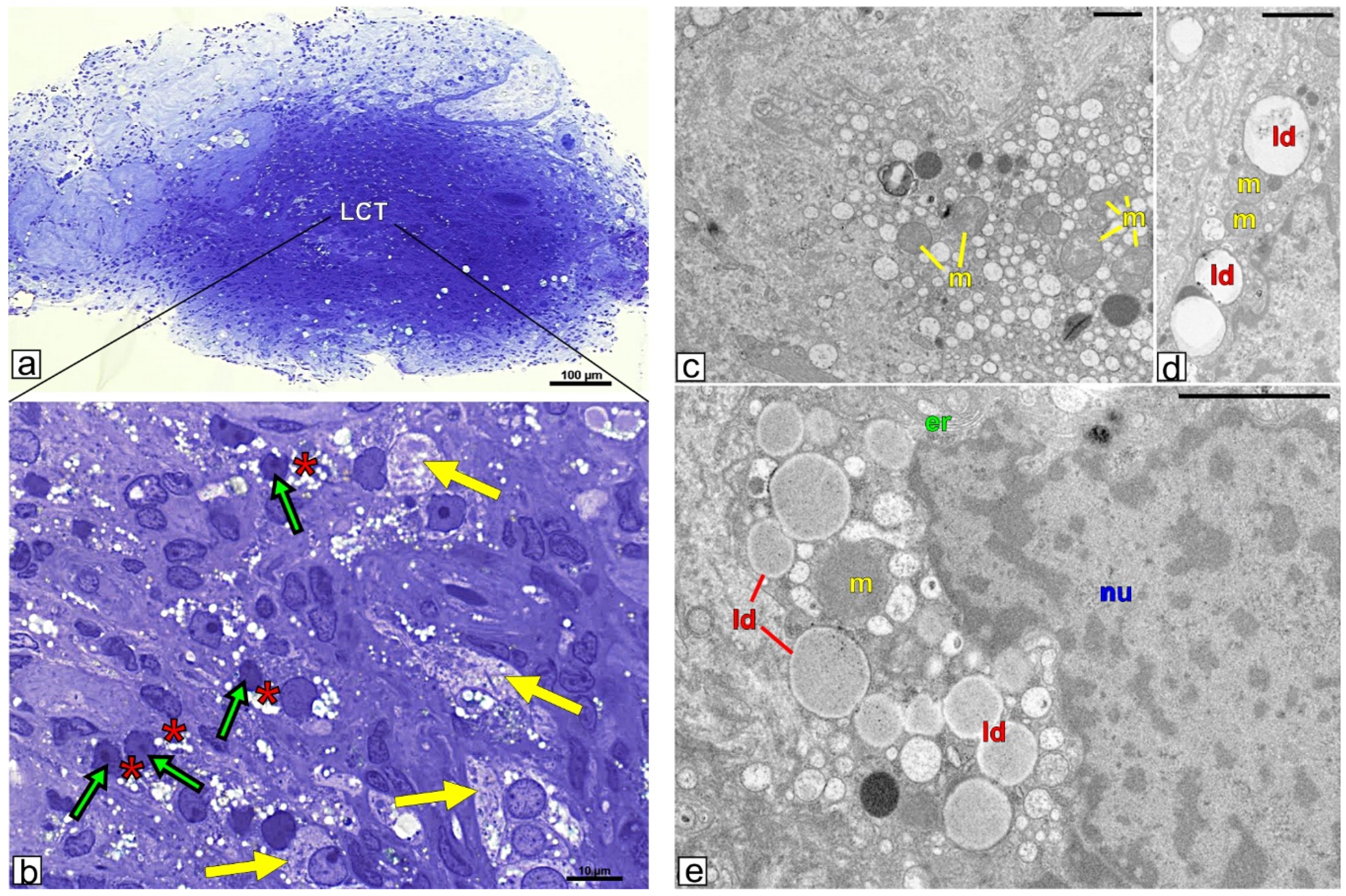
Morphology of Leydig cells in the testes after in vivo MCP-1 treatment.
Por um escritor misterioso
Descrição

Morphology of Leydig cells in the testes after in vivo MCP-1 treatment.

Therapeutic application of Sertoli cells for treatment of various diseases - ScienceDirect

Rapid Differentiation of Human Embryonic Stem Cells into Testosterone-Producing Leydig Cell-Like Cells In vitro
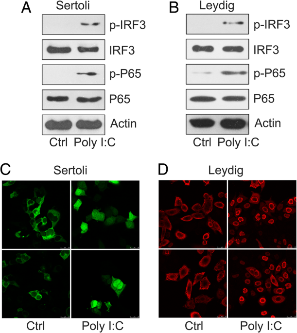
CircRNA-9119 suppresses poly I:C induced inflammation in Leydig and Sertoli cells via TLR3 and RIG-I signal pathways, Molecular Medicine
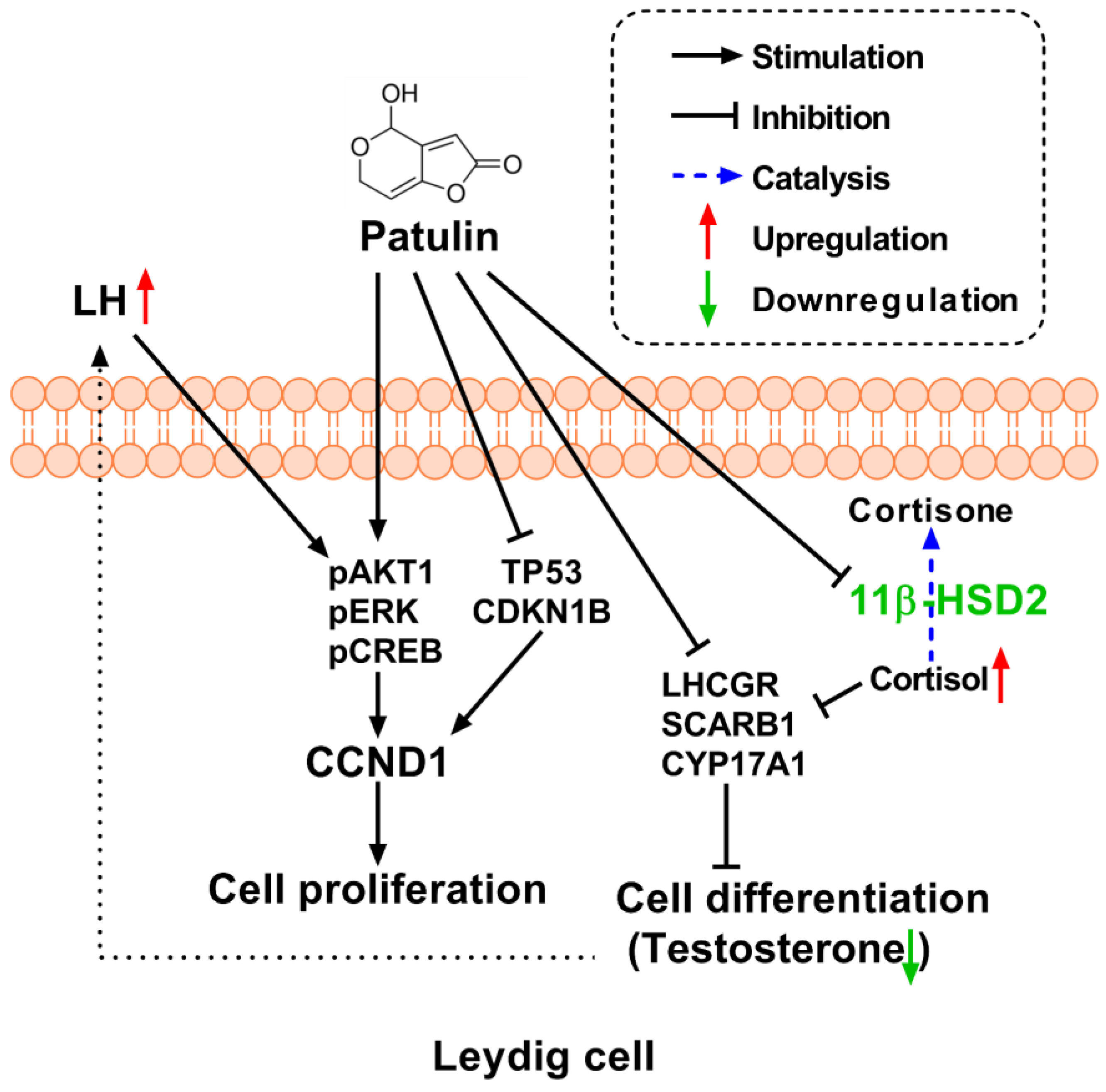
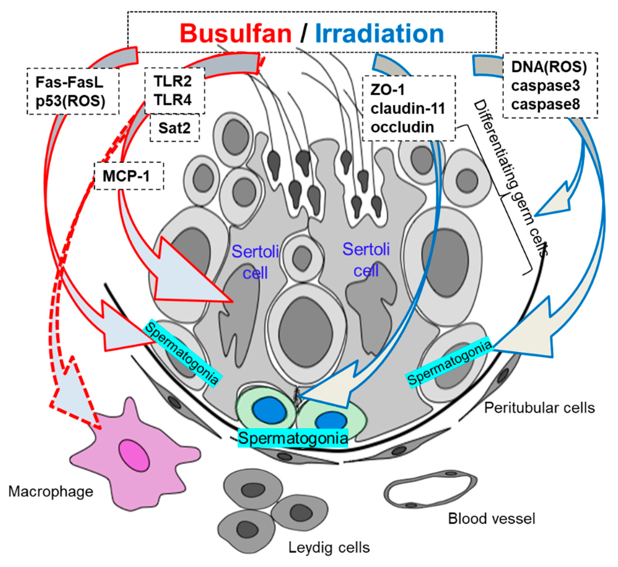
Toxins, Free Full-Text
IJMS, Free Full-Text

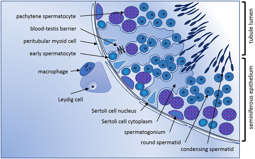
The Sertoli cell: one hundred fifty years of beauty and plasticity - França - 2016 - Andrology - Wiley Online Library

Insights into the Development of the Adult Leydig Cell Lineage from Stem Leydig Cells. - Abstract - Europe PMC
Cells, Free Full-Text
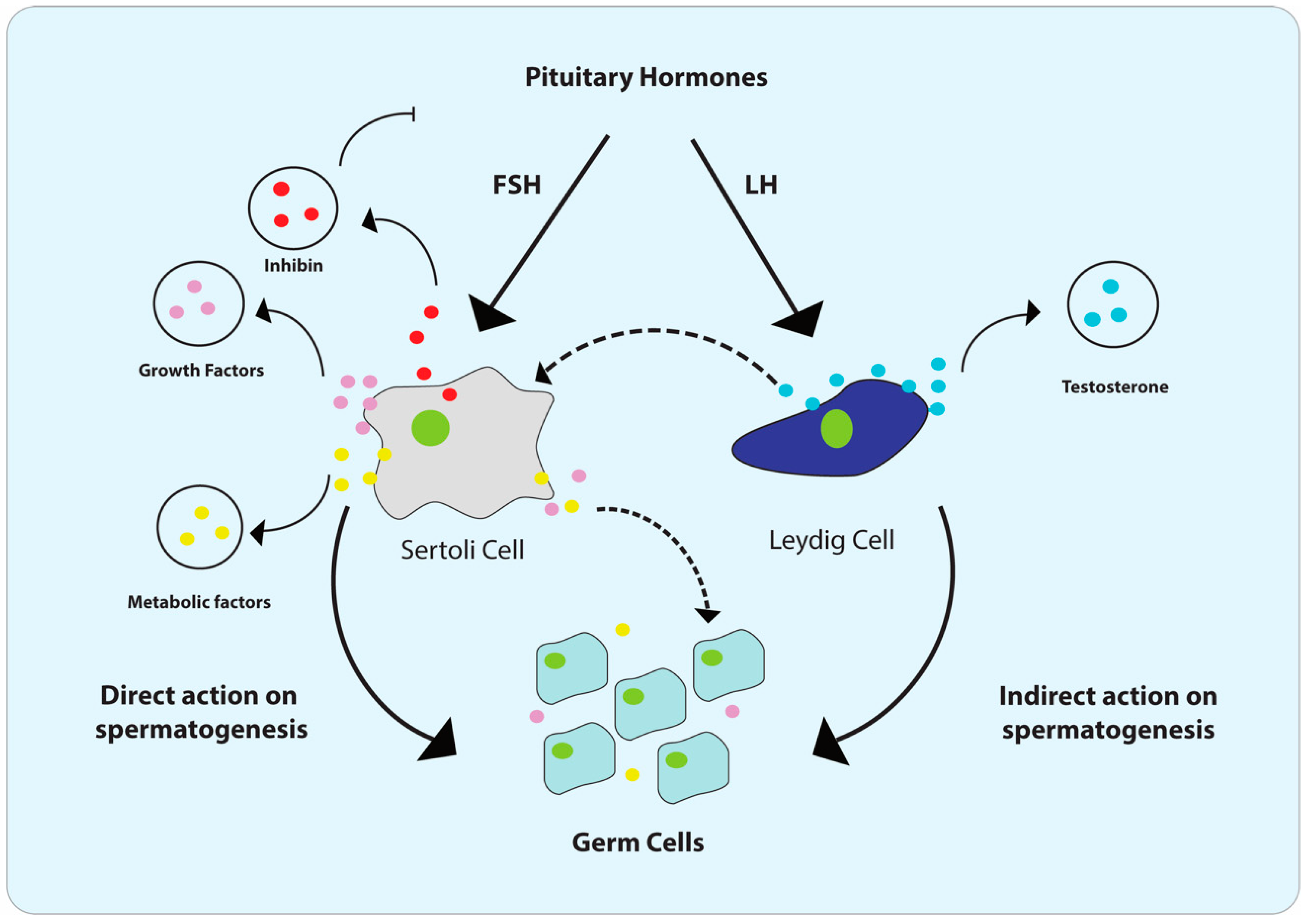
Frontiers Cytokines in Male Fertility and Reproductive Pathologies: Immunoregulation and Beyond
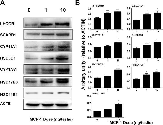
Monocyte Chemoattractant Protein-1 stimulates the differentiation of rat stem and progenitor Leydig cells during regeneration, BMC Developmental Biology

Stem Leydig cells: Current research and future prospects of regenerative medicine of male reproductive health - ScienceDirect
IJMS, Free Full-Text

Morphology of Leydig cells in the testes after in vivo MCP-1 treatment.

Transcription factor Dmrt1 triggers the SPRY1-NF-κB pathway to maintain testicular immune homeostasis and male fertility
de
por adulto (o preço varia de acordo com o tamanho do grupo)